
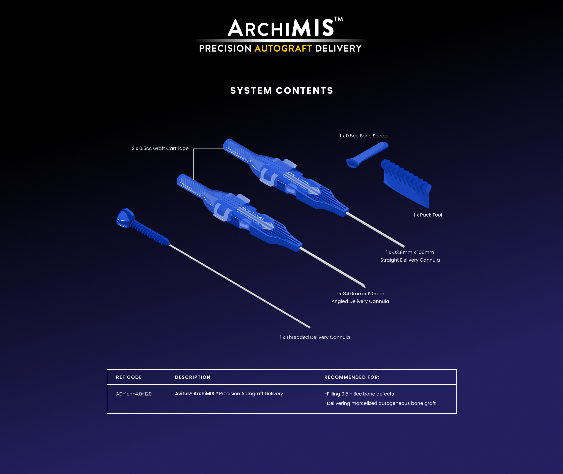
“The Avitus ArchiMIS™ system worked flawlessly in this case and is in alignment with the MIS wave of minimal invasive surgery. It makes sense from many aspects in regards to respecting the regional anatomy and its vasculature as well of the biological and mechanical components of healing. I would encourage all my colleagues in sports medicine, trauma surgery, foot & ankle and joint reconstruction fields to take a look at Avitus systems. It has become a great minimally invasive method that I’ve incorporated into my surgical armamentarium when bone graft and/or bone marrow harvesting is needed. I believe Avitus has helped improved the outcomes that I have been able to obtain in my patients; it is safe, easy to perform and utilizes the patient’s own biology to heal.”
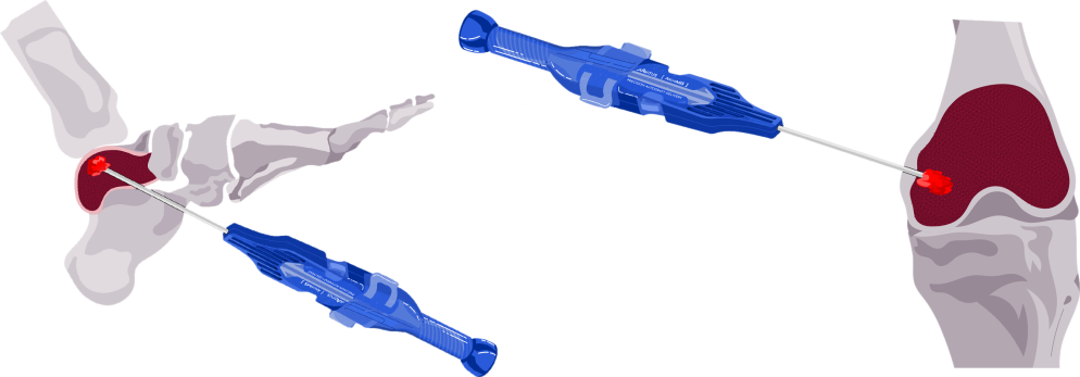
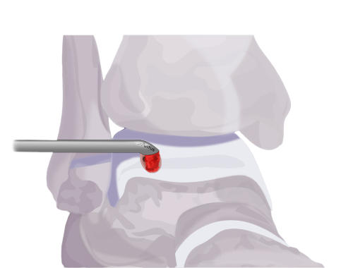
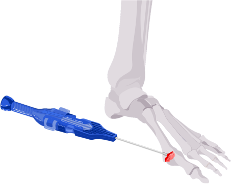
As a foot & ankle surgeon that regularly performs percutaneous and minimally invasive procedures; the development and launch of ArchiMIS™ provides me with an excellent option for precision autograft delivery. This innovative device fills an industry void by providing surgeons a first-of-its0kind solution for many foot & ankle pathologies that would otherwise need to be addressed through a traditional open approach, but now can be accomplished through MIS techniques.